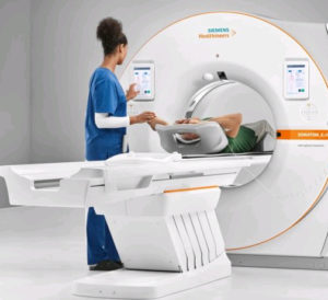
We have a powerful 64 slice CT scanner at our Sarasota center and a regular 16 slice CT at our Bradenton center.
 Both have very wide bores so claustrophobia is rarely ever an issue. These scanners allow for very fast scanning, up to 32 images per second. A chest or abdomen can be scanned in 10 seconds or less.
This results in a more comfortable exam for our patients with a faster scan and less time required for breath hold.
Beams of x-rays are passed from the scanner through the area of interest in the patient’s body from several different angles so as to create cross-sectional images, which then are assembled by computer into a three-dimensional picture of the area of the body that is being studied.
A CT (Computed Tomography) scan, often called a CAT scan, is a painless examination that gives your physician an unobstructed look at organs and structures that cannot be seen clearly on conventional X-rays. The scanner obtains image data from different angles around the body, and then uses computer processing of the information to show a cross-section of body tissues and organs.
The CT scan combines a sophisticated X-ray system with a high-speed computer. This combination produces a precise picture of the body, allowing the physician to see Tissue and Bone structure in fine detail. CT imaging is particularly useful because it can show several types of tissue (bone, blood vessels, lung, soft tissue) with great clarity. Radiologists can more easily diagnose problems such as cancers, cardiovascular disease, trauma and bone disorders.
CTA (Computed Tomography Angiography),
uses CT technology to visualize blood flow in arterial vessels throughout the body. Areas often studied include arteries serving the brain, the lung, the kidneys, the arms and the legs. Coronary calcium scores and Coronary CTA are examples of scans regularly performed. CTA can be used to detect narrowing or obstruction of blood vessels as well as aneurysms and embolism. CT and CTA can be combined with PET scans to create studies called PET Fusion scans.
 Low dose, CT lung cancer screening,
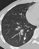
is an exam regularly performed. Lung Cancer is the leading cause of cancer-related deaths in the United States and represents about 25% of all cancer deaths. It is estimated that in the US, over 90 million individuals have a history of cigarette smoking, with approximately half reported to be current smokers. Stopping smoking reduces the risk for lung cancer; however, former smokers remain at an elevated risk compared to individuals who never smoked. Based on the National Lung Screening Trail (NLST), involving over 53,000 current and former heavy smokers, compared to standard chest x-ray, the mortality rate for lung cancer deaths was 20% lower in the group screened with low dose helical CT scans. The goal of early detection of lung cancer by using low dose CT is to find it when it’s most curable.
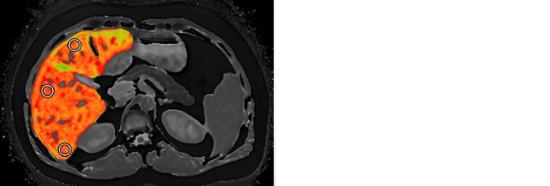
CT is one of the best tools for studying the chest and abdomen, and for diagnosing certain cancers including lung, liver and pancreatic. The image can allow a physician to confirm the presence of a tumor, and to measure its size, precise location and possible involvement with other tissues.


