


Partners Imaging Center has a Siemens PET/CT scanner at its Sarasota location
Combining PET and CT into one exam gives doctors two crucial pieces of information, function and anatomy, to help prescribe the best course of treatment for their patients. A CT scan provides detailed anatomy within the human body. Together, a PET/CT scan allows doctors to view metabolic activity and pinpoint where abnormal lesions are located so that they can target disease.
Positron Emission Tomography (PET) is a powerful diagnostic tool that often provides answers that no other imaging test can provide. PET is a non-invasive procedure. It helps physicians in the areas of Oncology, Cardiology and Neurology. Biochemical changes can be detected by a PET scan after a compound that contains radioactive molecules, bound to a sugar-like substance is injected into the body. These molecules provide the tracers that allow the measurements of metabolic activity within the body.
A computer records this information and converts it into pictures for diagnostic purposes. After a small amount of radioactive bound sugar is injected into the body, areas on the scan that show a high degree of metabolic activity have a greater chance of being cancerous.
 PET scans are used to detect cancer and to examine the effects of cancer therapy. These scans are performed on the whole body. PET scans of the heart can be used to determine blood flow to the heart muscle and help evaluate signs of coronary artery disease.
It can help determine if procedures such as angioplasty or coronary artery bypass surgery would be appropriate treatments for blood flow problems. PET scans of the brain are used to evaluate patients who have memory disorders of an undetermined cause; who have suspected or proved brain tumors; or who have seizure disorders that are not responsive to therapy, therefore, are candidates for surgery.
PET Imaging of Alzheimer’s and other Dementias
Early Alzheimers Disease (AD) is often difficult to diagnose clinically. PET imaging using (F-18) FDG has been shown to be accurate in the early detection of AD. The Centers for Medicare and Medicaid Services have approved FDG-PET imaging as a routine exam for the early and differential diagnosis of AD, specifically to differentiate from Frontotemporal Dementia (FTD). This is based on evidence showing that adding PET to a clinical exam increases diagnostic sensitivity in AD.
How does FDG-PET/CT work? One of the earliest changes in AD is altered glucose metabolism. Some researchers are now referring to AD as Type 3 Diabetes because of relative insulin resistance in the brain of AD patients resulting in less glucose uptake. FDG-PET measures cerebral metabolic rate of glucose uptake. By the time a patient has dementia symptoms, a reduction in glucose uptake has already occurred and can be measured by PET. In fact, FDG-PET is so sensitive to glucose uptake, that patients with increased risk for AD (e.g. family history or genetic susceptibility) will often show reduction in glucose uptake within specific areas of the brain before the onset of clinical symptoms.
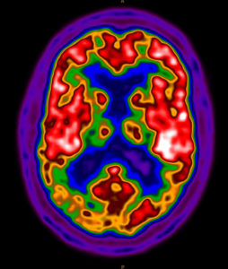
 What does AD look like on PET scan?
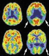
The FDG-PET pattern of early AD is very specific. It shows a pattern of abnormally low glucose uptake (hypometabolism) in specific areas of the brain, the posterior cingulate, precuneus, and temperoparietal regions. This can be observed on visual inspection.
In addition, at Partners Imaging, we create topographic maps of the brain that quantitate the degree of hypometabolism in each region. Also, the degree of glucose uptake in each region is compared to an age-matched control group of cognitively normal individuals to give us a “z-score” or statistical measure relative to normal glucose uptake
